


The Newfoundland Club
Promoting Heart Testing in Newfoundlands
Understanding Heart Testing
by Dr. Jo Dukes McEwan BVMS, MVM, PhD, DVC, MRCVS
Both the original and the new heart test certificates have led to some confusion in people’s minds because of the difficulty in understanding the results. The new certificates have solved problems in having separate copies for the owner, the dog’s vet, the cardiologist and research collation, and they are less onerous to fill out.
The heart test result is given as the Grade of any heart murmur detected by the cardiologist. Murmurs are graded as 1 to 6 out of 6. Very quiet murmurs are graded 1/6 and the loudest murmur is a 6/6. The Veterinary Cardiovascular Society have established some guidelines for UK cardiologists to ensure that grading of murmurs detected with a stethoscope is as consistent as possible between cardiologists.
If no heart murmur is detected, then a grade 0/6 can be inferred. The results of the echo-Doppler examination (if done) should be checked before it can be assumed that the dog is normal.
HEART MURMURS MAY OCCUR FOR VARIOUS REASONS
Heart murmurs may be due to:
- Narrowed valves (offering increased resistance to flow, so flow velocity (speed) has to increase to get through the obstruction. Think of a narrow part of a river compared to a wider part. The flow is fast and turbulent in the narrow part. This results in a heart murmur). Examples are aortic or sub-aortic stenosis or pulmonic stenosis.
- Leaky valves. As a leak of blood jets through the incompetent valve, it results in a murmur, since the abnormal flow (regurgitation) is fast and turbulent. Most murmurs associated with leaky valves are due to mitral or tricuspid regurgitation.
- Abnormal shunts. If there is a defect in the heart, such as a ventricular septal defect (VSD; a hole between the left and right ventricles) or a Patent Ductus Arteriousus (PDA), blood passes through these areas resulting in a heart murmur.
| Normal laminar blood flow |  Turbulent, high velocity blood flow after the obstruction Turbulent, high velocity blood flow after the obstruction |
|
|
Figure 1. Illustrating how blood flow passing a narrowed area (e.g. stenotic or leaky valve) results in change from normal (laminar) flow to turbulent flow. The high velocity turbulent flow in the heart may result in an audible heart murmur. |
||
Sub-aortic stenosis is the main congenital heart disease of concern in Newfoundland dogs. Confirmation of diagnosis by echo-Doppler is based on a Doppler recording of a peak aortic velocity exceeding 1.7 metres per second in a relaxed dog. The blood flow through the aortic valve is normally much less than 1.7 m/s. Increased velocity is associated with increased turbulence of blood. These are both factors in creating a heart murmur. The abnormal flow is a consequence of a narrowed (stenotic) area affecting the aortic valve or caused by a fibrous ridge below the valve.
There are other congenital heart diseases occasionally seen in Newfoundlands, which may be associated with a murmur, or in some cases (such as severe tricuspid valve dysplasia) no murmur is detected. Murmurs may be continuous such as in patent ductus arteriosus (PDA).
In DILATED CARDIOMYOPATHY (DCM), there is no primary defect affecting the heart valves or flow of blood through the heart, and so NO heart murmur is often recorded (0/6). This must NOT be interpreted as the dog having a normal heart. The heart test result has to be interpreted based on the echo-Doppler result. Often, an abnormal heart rhythm is detected, with abnormal beats (“ectopics”) or atrial fibrillation – before any murmur. Later in the course of DCM, since the chambers of the heart become massively dilated, the mitral valve annulus (the “scaffolding” supporting the leaflets of the mitral valve) becomes stretched, and this can have the consequence of mitral regurgitation – which is then detected as a heart murmur. In DCM, therefore, in the early stages, there may be NO abnormalities listening through a stethoscope but as the disease develops, murmurs will develop, although rarely higher than grade 2/6. Other abnormal findings can be detected with a stethoscope in dogs with heart failure, prior to successful treatment. We can hear “gallops” which are due to the stiffened ventricles filling with blood. Gallops are always abnormal. It is satisfying to monitor these during treatment. Once heart failure is controlled, the gallops resolve.
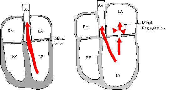
Diagram illustrating a normal heart (left) and the changes seen with DCM (right). The four chambers of the heart are labelled; right atrium (RA), right ventricle (RV), left atrium (LA) and left ventricle (LV). The mitral valve is situated between the left atrium and left ventricle. Normally, during the relaxed stage of the heart cycle (diastole), blood flows from the left atrium to the left ventricle through an open mitral valve. During ventricular contraction (systole), blood flows out of the LV into the aorta and the mitral valve closes (large arrow, left). In DCM (right), the chambers of the heart are dilated and the walls are thin. The mitral valve annulus is “stretched”, so during ventricular contraction, as well as normal blood flow into the aorta, some flows retrograde across the valve leafets into the left atrium, giving rise to turbulent blood flow which may be heard as an audible heart murmur. This explains why some dogs with DCM have a heart murmur graded on heart testing.
THE GRADE OF A HEART MURMUR DOES NOT NECESSARILY CORRELATE WITH THE SEVERITY OF HEART DISEASE
OTHER FACTORS INFLUENCE HEART MURMURS
In SAS, there may be NO murmur in a young pup. As the pup grows and the heart grows, the abnormality below the valve remains constant, so it becomes proportionately worse with ageing. This is the reason that dogs should be over 12 months old for the results of official heart testing to be recorded or published. Although it is sensible to get pups checked, they must be re-examined at over 12 months old. There is a possibility that SAS does become more severe up until 24 months of age, but then remains reasonably constant.
We have shown that some Newfoundlands, being broad-chested, possibly over-weight, and tending to pant, have NO audible heart murmur. There is evidence of SAS on echo-Doppler. This shows that the sensitivity of heart testing by the stethoscope alone is insufficient to detect all cases of SAS (although it will certainly detect the more severe cases). This is the reason why dogs used at stud should be echo-Dopplered for SAS. Stud dogs are potentially responsible for many more progeny than bitches.
Dogs which had aheart murmur detected at 12 months old will mature and may put on a lot of weight and then the murmur becomes undetectable. Similarly, dogs with no heart murmur detected may lose a lot of weight, and then one becomes apparent.
In DCM there may be no murmur, despite the fact mitral regurgitation is confirmed on echo-Doppler. This is because the contractility of the heart may be so reduced that the heart muscle does not generate sufficient force for the jet of blood (of mitral regurgitation) to be fast enough to result in an audible heart murmur.
ECHO-DOPPLER SCREENING FOR DCM
DCM is an acquired heart disease. The dog is not born with it. It gradually develops after adult-hood and the average age of symptoms developing is 8 years old. If we scan an affected dog about 12 – 24 months prior to the development of symptoms of breathlessness, exercise intolerance or coughing, the classical features of DCM are evident on echocardiography. These features are:
- Dilated, rounded left ventricle.
- Walls of the ventricles appear thin.
- Severely reduced contractility of the left ventricle. The fractional shortening is an easy-to-obtain indicator of contractility. In DCM, it is less than 22%.
- Dilated left atrium
- May have arrhythmias such as atrial fibrillation detected on the ECG through the scan.
Because these dogs show no symptoms, we call this OCCULT DCM. Even if no heart murmur is detected (0/6), these dogs are ABNORMAL and have significant heart disease. This will be shown in the echo-Doppler section of the heart test form. It is not possible to predict how rapidly the disease will progress to eventually result in symptoms. In some dogs, it may be rapid and in others, it may take over two or more years. The older a dog is at the time of diagnosis of occult DCM, the more slow the progression tends to be. The progression tends to be faster in dogs compared with bitches.
Prior to this, we may identify other echo-Doppler abnormalities in Newfoundlands. These are:
-
LEFT VENTRICULAR ENLARGEMENT (LVE)
Only occasional Newfies have this abnormality, but they do progress to develop more classical DCM within 12 – 18 months, although it is often longer before they show symptoms. Male dogs with a LV dimension of over 55 millimetres and bitches of over 50 mm are abnormal. These dogs have a normal fractional shortening.
-
DEPRESSED FRACTIONAL SHORTENING (dFS)
These Newfies have a normal elliptical shape to the left ventricle and it is not dilated. There only abnormality is a very low fractional shortening measurement (and other parameters of contractility also confirm the pumping ability of the LV is impaired). Fractional shortening less than 18% is very low. Between 18 and 20% may be equivocal.
In the four years or so that we have been monitoring Newfies with severe dFS (less than 18%), only 5 out of 29 dogs developed DCM. Most of the dogs have remained similar on repeat scanning. There are still unanswered questions about this group:
- Will these dogs go on to develop DCM in the future?
- Do they have a limited manifestation of DCM which will not progress?
- Is it a normal variation? (This is unlikely, since we do not see this in Newfies where there is no positive family history of DCM).
Only by continuing the serial scans through the dogs' lives will we be able to answer these questions.
A note will be made in the heart test forms if these EQUIVOCAL findings are identified.
SERIAL SCANS ARE REQUIRED TO EXCLUDE DCM
DCM: THE GENETIC STUDIES
It is vital that I know what happens to dogs with echo-Doppler equivocal findings, for the genetic analysis. Although I have a DNA sample from most of the dogs I have scanned, I need to be certain that dogs are definitely normal or abnormal by the end of their lives. If we obtain this information, by serial scans or by examining hearts obtained at post-mortems, it will help the genetic studies – and I am extremely grateful to all Newfie owners and breeders who have allowed the serial scans. With this confirmed information, the genetic studies should result in a blood test for DCM which can be used in young dogs, which will identify dogs at risk for developing DCM.